
Advertisement
California-based researchers announced that they found a much faster way to scan the 3D structures of certain small molecules. Their improved approach provides incredibly clear images of objects the size of certain hormones within a few hours or even just 30 minutes.
They took an existing technique called micro-electron diffraction (MicroED) and adjusted it to work on molecules that are smaller than the proteins it usually targets. Their study showed that not only did the adaptation work on small molecules, it also took a much shorter amount of time to set up.
Whereas conventional MicroED scans needs the molecules in the starting sample to match the size of salt grains, the modified method does not need any special preparation. It will even work on powdered scrapings from the beaker.
“We took the lowest-brow samples you can get and obtained the highest-quality structures in barely any time,” remarked California Institute of Technology (CalTech) researcher Brian Stoltz. “When I first saw the results, my jaw hit the floor.” (Related: These highly effective alternatives to toxic medication help beat depression.)
Micro-electron diffraction can view small molecules thanks to the latter being crystals
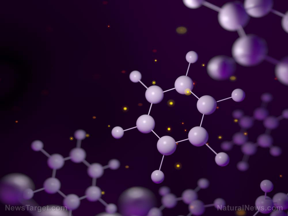
To the naked eye, a sample of small molecules appears to be made of fine but ordinary powder. But with the right scanning equipment, the “powder” reveals itself to be a collection of microcrystals, a billion of which can fit inside a piece of dust.
Researchers previously believed that they could only view the molecular structures of these extremely tiny crystals with great difficulty. They also didn’t think micro-electron diffraction scans could do the job. Now, thanks to the efforts of CalTech researchers and their counterparts at the University of California Los Angeles (UCLA), the modified MicroED can be used to observe the crystalline structures of small molecules.

A molecule counts as “small” if it weighs less than 900 daltons, one of which is equal to an atom of hydrogen. The weight classification includes the likes of natural chemical compounds, hormones and certain other biological compounds, and some pharmaceutical drugs.
The modified MicroED methodology makes it fast and easy to view the structures of these microcrystals. It can be used to create new chemical compounds, analyze minuscule pieces of evidence recovered from crime scenes, and performing a bevy of accurate medical tests.
From drug testing to putting together small molecules to form new compounds
Micro-electron diffraction can analyze blood samples of athletes for signs of performance-enhancing drugs. These illegal drugs are difficult to detect because they barely leave a chemical trace. But MicroED scanning can confirm the absence or presence of steroids and other associated small molecules.
Another role is to let researchers view the microcrystals during the intermediate and last parts of a chemical reaction. These are the most important parts of assembling a chemical compound. An error committed during either segment would ruin the effort, so a chemist needs to review the intermediate and final molecules formed by the reaction.
MicroED works by showering electrons upon a chemical sample. As the electrons diffract off the atoms of the chemical, they create a pattern that matches the three-dimensional structure of the crystal.
Normally, the methodology requires big crystallized proteins that give off clear diffraction patterns. Then UCLA researcher Tamir Gonen – the co-author of the study along with CalTech’s Stoltz – mentioned that powdery samples sometimes sufficed for the finicky technique to work.
Upon remembering that the powder was made up of microcrystals, the authors realized that they could skip the crystallization process and scan the sample using MicroED. A series of tests proved that they could get crystal clear images of the structures of small molecules, and they hope their technique will enter wide use.
Sources include:
Submit a correction >>
This article may contain statements that reflect the opinion of the author
Advertisement

Advertisements


NewsTarget.com © 2021 All Rights Reserved. All content posted on this site is commentary or opinion and is protected under Free Speech. NewsTarget.com is not responsible for content written by contributing authors. The information on this site is provided for educational and entertainment purposes only. It is not intended as a substitute for professional advice of any kind. NewsTarget.com assumes no responsibility for the use or misuse of this material. Your use of this website indicates your agreement to these terms and those published on this site. All trademarks, registered trademarks and servicemarks mentioned on this site are the property of their respective owners.
