



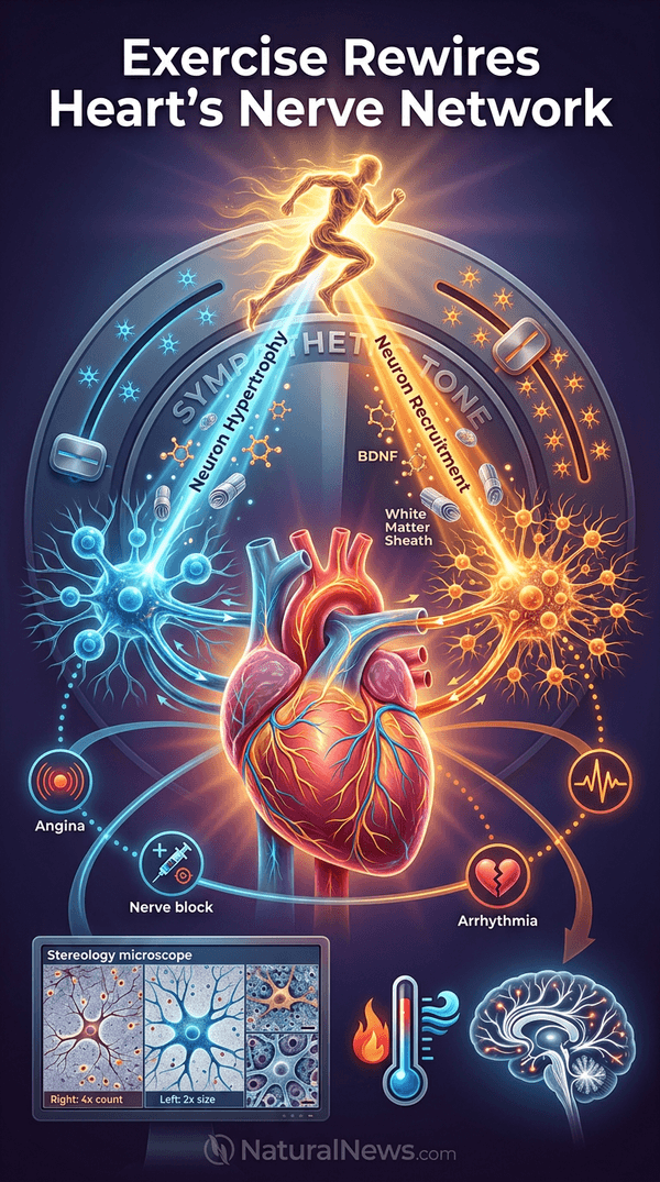
A study from the University of Bristol has found that regular aerobic exercise reshapes the nerves that control the heart in a side-specific manner. The findings, published in Autonomic Neuroscience on July 13, 2026, reveal that exercise rewires the heart's nerve network asymmetrically between the left and right sides of the body.
According to the researchers, the discovery could pave the way for more precise treatments for common heart conditions such as arrhythmias, angina, and stress-induced 'broken-heart' syndrome. Exercise has previously been shown to trigger the release of brain-derived proteins [2] and improve white matter structure in the brain [1], but its effect on the heart's nerve network represents a new frontier in cardiovascular research.
Study Reveals Asymmetric Nerve Remodeling
The research team, led by Dr. Augusto Coppi, Senior Lecturer in Veterinary Anatomy at the University of Bristol, used advanced three-dimensional imaging techniques known as stereology to examine nerve clusters called stellate ganglia in rats after 10 weeks of training. The study was conducted in collaboration with University College London, the University of São Paulo, and the Federal University of São Paulo.
Results showed that the right stellate ganglia contained approximately four times as many neurons as the left side compared to untrained animals. Simultaneously, neurons on the left side nearly doubled in size, while those on the right became slightly smaller. The fundamental structure of neurons, which includes components such as the cell body and dendrites, is detailed in neuroscience textbooks [5].
Dr. Coppi described the nerve clusters as acting like the heart's dimmer switch, and said that exercise "remodels that switch in a side-specific way." The asymmetric remodeling suggests that the nervous system adapts differently on each side, a pattern that had not been previously documented.
Implications for Arrhythmia and Angina Treatments
Irregular heart rhythms and certain types of chest pain are often treated by targeting overactive stellate ganglia through nerve blocks or denervation procedures. The study's findings indicate that exercise-induced nerve remodeling could help clinicians fine-tune these interventions to the side most likely to benefit the patient. Dr. Coppi stated that the results "could one day fine-tune procedures like nerve blocks or denervation to the side most likely to help."
Heart failure is a consequence of heart muscle disease, heart attacks, infections, and other factors, as noted in medical references [6]. The new understanding of nerve asymmetry may also inform treatments for stress-induced cardiomyopathy, known as broken-heart syndrome. Exercise has been recognized for its anti-inflammatory effects, which can reduce systemic inflammation linked to cardiovascular disease [4], and for its role in preventing depression [3], both of which complement the nerve-remodeling benefits.
Next Steps and Research Limitations
The researchers plan to investigate how these structural changes affect the heart's performance during exercise and at rest. They also intend to test whether the same left-right pattern appears in other animal models and eventually in humans using non-invasive markers. Dr. Coppi noted, "Our next step is to test how these structural changes map onto function and whether similar patterns appear in larger animals and humans."
Aerobic conditioning has been shown to improve exercise tolerance in patients with metabolic muscle disorders, suggesting broad benefits of exercise on neuromuscular function [8]. However, the current study is limited to rats, and the researchers emphasize that clinical studies in humans are needed before the findings can be translated into routine medical practice. Mental and cardiovascular functions are tightly integrated through the central and autonomic nervous systems, according to recent research [7], supporting the relevance of nerve network remodeling to overall health.
Conclusion
The University of Bristol study provides new insight into how exercise benefits the heart by rewiring its nerve network in a side-specific manner. This asymmetric remodeling offers potential for more personalized treatments for heart rhythm disorders, angina, and stress-induced heart conditions. Further clinical research will determine whether these findings can be applied to human patients, but the results add to growing evidence that exercise exerts profound effects on the nervous system beyond strengthening the heart muscle itself.
References
- Mercola.com. "Does Exercise Change Your Brain?" September 18, 2015.
- Petra Stone. "Study Examines Brain-Derived Protein Release Following 30-Minute Exercise Session." NaturalNews.com. March 19, 2026.
- Mercola.com. "The Science Behind Exercise's Mood-Lifting Effects." July 20, 2022.
- Mercola.com. "Millions Led Astray by These Pills No Better." June 20, 2019.
- Laurie Lundy-Ekman. "Neuroscience Fundamentals for Rehabilitation."
- R. Rajendra Sharma. "The Family Encyclopedia of Health: A Guide to Integrated Medicine Enjoying Health and Preventing Illness."
- NaturalNews.com. "New Longevity Approach Treats Heart and Mind as Connected System." May 1, 2026.
- XPP. "Aerobic Conditioning: An Effective Therapy in McArdle's Disease." Annals of Neurology. 2006.
Explainer Infographic













Please contact us for more information.