



Exercise Alters Heart-Controlling Nerves
Regular aerobic exercise reshapes the nerves that regulate the heart, according to a July 2026 study from the University of Bristol. The study, published in Autonomic Neuroscience, found that after 10 weeks of training, rats showed asymmetric changes in the stellate ganglia nerve clusters -- paired structures in the lower neck and upper chest that send “go faster” signals to the heart.
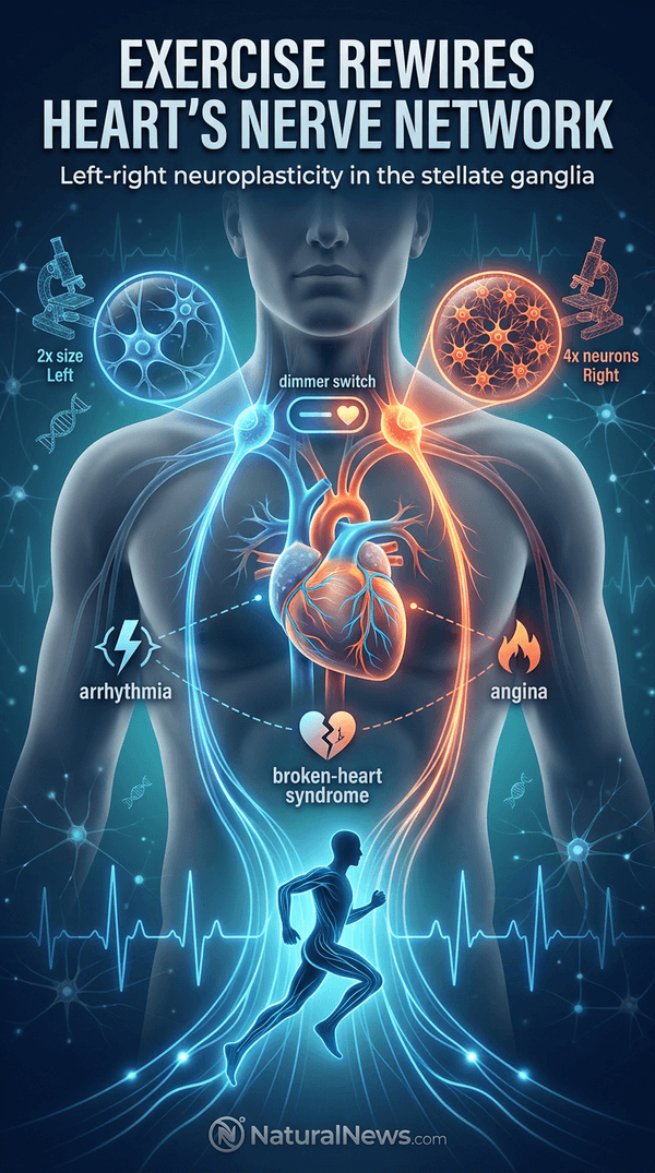
Lead author Dr. Augusto Coppi, Senior Lecturer in Veterinary Anatomy at the University of Bristol, said that the discovery reveals a previously hidden left-right pattern in the body’s “autopilot” system for the heart. [1] This finding could eventually help doctors tailor treatments for arrhythmias, angina, and stress-induced “broken-heart” syndrome, according to the researchers.
Key Findings: Side-Specific Nerve Remodeling
Using advanced three-dimensional imaging techniques called stereology, the team examined nerve cluster changes after the exercise regimen. Trained rats had about four times as many neurons in the right cardiovascular nerve cluster compared with the left, relative to untrained animals. [1] At the same time, neurons on the left side nearly doubled in size, while those on the right became slightly smaller, the researchers reported.
Dr. Coppi described the nerve clusters as acting like “the heart’s dimmer switch,” noting that exercise remodels that switch in a side-specific way. The stellate ganglia are part of the sympathetic chain, which regulates heart rate and contraction force, as described in functional anatomy texts. [2] The asymmetric changes suggest that aerobic training induces a previously unrecognized form of left-right neuroplasticity in the autonomic nervous system.
Potential Clinical Implications
The findings may inform treatments for arrhythmias, stress-induced “broken-heart” syndrome, and angina, which are often addressed by targeting the stellate ganglia. According to researchers, the left-right differences could help fine-tune procedures such as nerve blocks or denervation to the side most likely to provide benefit. [1] The study offers clues that could one day allow doctors to target therapies more precisely, though clinical trials would be needed, Dr. Coppi said.
Exercise has long been known to improve cardiovascular health; for example, resistance-based interval training has been shown to improve endothelial function in people with Type 2 diabetes. [3] The new study extends this understanding by showing that exercise not only strengthens the heart muscle but also rewires its neural control system.
Study Limitations and Next Steps
The research was conducted in rats and is considered early-stage; the authors cautioned that clinical application requires further investigation. [1] Planned next steps include testing how the structural changes affect heart performance during exercise and at rest, using non-invasive markers in humans.
“Understanding these left-right differences could help us personalize treatments for heart rhythm disorders and angina,” Dr. Coppi said in the statement. The team also aims to determine whether the same left-right pattern appears in other animal models and ultimately in human patients, according to the study.
Conclusion: Foundational Research for Targeted Therapies
The study provides the first evidence that aerobic exercise induces asymmetric neuroplasticity in the stellate ganglia, a key component of the autonomic nervous system that helps regulate circulation, respiration, and metabolism. [4] Researchers stated that understanding these left-right differences could lead to personalized treatments for heart rhythm disorders and difficult-to-treat angina. The work was supported by multiple institutions, including University College London, the University of São Paulo, and the Federal University of São Paulo in Brazil, and references are available in the journal Autonomic Neuroscience.
References
- Exercise Rewires Heart’s Nerve Network, Study Finds - NaturalNews.com. July 15, 2026.
- William O. Reece. "Functional Anatomy and Physiology of Domestic Animals".
- Diabetic patients can improve the function of their blood vessels by doing resistance-based interval exercise - NaturalNews.com. June 08, 2018.
- Laurie Lundy-Ekman. "Neuroscience fundamentals for rehabilitation".
- THE EFFECTS OF THERAPEUTIC ULTRASOUND ON HEART RATE VARIABILITY: A PLACEBO CONTROLLED TRIAL - Journal of Ultrasound in Medicine. 2005.
Explainer Infographic








Please contact us for more information.