


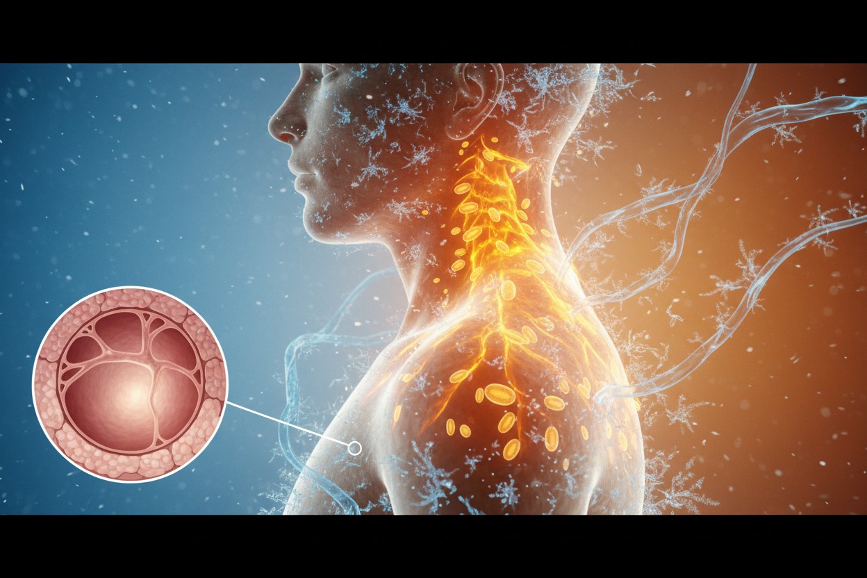
- Brown fat burns calories via thermogenesis, unlike white fat which stores them.
- New research identifies SLIT3 protein fragments that build brown fat’s blood vessel and nerve networks.
- Without these networks, brown fat cannot function effectively for metabolism.
- People with obesity tend to have less brown fat infrastructure, reducing calorie-burning ability.
- New research opens the door to potential therapies that boost natural fat-burning capacity rather than suppress appetite.
What this means for metabolism
For decades, scientists have known that brown adipose tissue—unlike its white counterpart—burns calories rather than storing them. But a fundamental question remained unresolved: What allows brown fat to function effectively? New research published Nov. 11 in Nature Communications provides an answer. Investigators at multiple institutions analyzed fat tissue samples from more than 1,500 individuals, including those with obesity, to uncover how brown fat develops the blood vessel and nerve networks essential for its calorie-burning capacity. The findings reveal a protein system that could fundamentally shift approaches to obesity treatment.
The two-part protein system that powers brown fat
Brown fat generates heat through thermogenesis, a process requiring both blood vessels to deliver oxygen and nutrients and nerves to signal activation. The study identified a protein called SLIT3, secreted in high amounts by brown fat cells, that splits into two distinct fragments with separate functions. SLIT3-N promotes blood vessel growth, while SLIT3-C builds nerve networks. Both fragments must work together for brown fat to burn calories effectively.
The researchers discovered that adipocyte progenitors—precursor cells to fat cells—secrete SLIT3, which is then cleaved into these functionally distinct fragments. The study also identified PLXNA1 as a receptor for SLIT3-C and BMP1 as the first SLIT protease described in vertebrates.
Why infrastructure matters for metabolic health
People with obesity tended to have less of this brown fat infrastructure, which may explain why their brown fat is often less active. The finding challenges the conventional view that obesity stems solely from calorie imbalance. Instead, it suggests that the body’s ability to burn fat depends on cellular infrastructure that can be compromised.
The study builds on earlier work published in Cell in 2014, which identified the transcription factor IRF4 as a key driver of brown fat thermogenesis. That research established that IRF4 interacts with PGC-1-alpha to promote UCP1 expression, the protein that allows brown fat cells to dissipate energy as heat. The current study extends this understanding by revealing how the tissue network supporting UCP1 function is built.
Current treatment models offer an incomplete approach
The conventional calories in, calories out” model has dominated obesity research for a century despite limited progress. A 2023 Nature review of 100 years of data found that people’s basal metabolic rates have been plunging over the last 30 years, suggesting the problem runs deeper than simple energy balance. The pharmaceutical industry’s response—drugs like semaglutide that suppress appetite—addresses only one side of the energy equation while generating new health risks.
The discovery of SLIT3’s role opens a different path: boosting the body’s natural ability to burn calories rather than fighting appetite. This approach aligns with a growing understanding that metabolic health depends on the body’s internal systems functioning as nature intended.
Natural support for brown fat activity
While therapies based on this research remain years away, some strategies may support existing brown fat function:
- Cold exposure through brief cold showers or cooler environments
- Regular exercise linked to improved brown fat function
- Foods containing capsaicin from chili peppers or compounds in green tea
These approaches support existing brown fat activity and the development of the necessary infrastructure, but do not build the new blood vessel and nerve infrastructure that the SLIT3 system creates.
The path forward
This research reveals that brown fat requires more than mere existence to burn calories—it needs a complete support system. The coordinated neurovascular expansion mediated by distinct SLIT3 fragments provides a bifurcated yet integrated mechanism ensuring synchronized brown adipose tissue response to environmental challenges. For now, practical applications remain on the horizon, but the discovery offers a promising direction for treatments that help the body become better at burning fuel, working with natural metabolism rather than against it.
Sources for this article include:











Please contact us for more information.